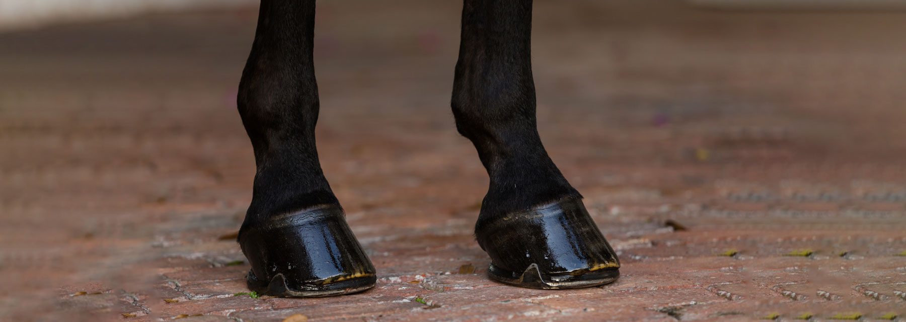
Top Farriers Weigh In On Concerns Affecting The Equine Hoof
Hoof Cracks
There is a wide spectrum of hoof cracks and the severity of the crack depends on location, depth and site of origin. Grass cracks and sand cracks tend to be superficial flaws caused by the environment. Heel cracks, bar cracks and toe cracks are capable of causing extreme pain. Quarter cracks, in particular, are painful, difficult to manage and have a variety of causes. Quarter cracks involve the coronary band and grow downward. They are more likely than other types of cracks to bleed and become infected, leading to extreme pain and lameness. For all cracks, the farrier should be involved in assessment and will determine the best plan for balance, stabilization and repair.
Wesley Meyer: “Nutrition, balance and concussion are three reasons for cracks. If the hoof is not getting enough good nutrition, it can fail. Secondly, watch that they’re balanced. See how the horse is landing when you watch them move. Is it concussion? Is it workload? If they’re balanced, but they come down a little off, the hooves can fail and result in cracks and other issues.”
Ian Curry: “Shoe for balance. That’s the most important thing and to stabilize the crack. It has to remain clean while the crack is still alive, and it has to be flushed out with an antibacterial solution. Then, move forward. As long as you can get the crack stable, you can send them back to work.”
Michael Majors: “It is crucial to clean and trim the crack often and also important to trim back the crack to the very end, so it does not continue to crack farther up the wall. I recommend a good supplement here to support hoof growth because the faster the feet grow, the sooner cracks will grow out too.”
Corey Payne: “The first thing I do when addressing hoof cracks is to determine the cause. Most of time, the crack is there because of excess load in that part of the foot. If the foot is not balanced to the horse’s confirmation, that is when cracks can develop. The ideal hoof is designed to bear weight evenly across its surface, so when the foot is out of balance and bearing too much weight on a certain part of the hoof, cracks can develop.”
“Nutrition, balance and concussion are three reasons for cracks. If the hoof is not getting enough good nutrition, it can fail. Secondly, watch that they’re balanced. See how the horse is landing when you watch them move. Is it concussion? Is it workload? If they’re balanced, but they come down a little off, the hooves can fail and result in cracks and other issues.”
— Wesley Meyer
Hoof Abscess
Caused by bacteria getting trapped inside the hoof, abscesses are a very common cause of acute lameness in horses. Trauma to the hoof sole caused by a puncture (e.g., nails, glass, etc.) may lead to bacteria and debris having access to the inner hoof and resulting in an abscess. Excess moisture may cause the hoof wall to soften and allow bacteria access through gaps in the white line. Conversely, extremely dry hooves may lead to brittleness and cracking of the hoof wall, which can also lend access to bacteria. The hoof itself does not allow room for swelling. So, as excess pressure builds up from foreign bacteria, it causes an extreme amount of pain that results in severe lameness that often seems to occur overnight. In most circumstances, a farrier or veterinarian will be able to drain the abscess, with pain relief being nearly immediate, and allow healing to begin.
Payne: “When treating abscesses, I try to establish a drain track in the bottom of the hoof, if possible. This helps relieve the pressure that is causing the horse pain and allows all the infected material a way to exit the hoof. On every horse with a suspected abscess, the use of hoof testers is the best way to attempt to localize the point on the sole that exhibits the most pain. If it’s still unclear where the abscess is located, I will utilize foot radiographs to look for gas in the hoof capsule. The bacteria in the abscess produce gas that will show up on an X-ray and help determine where the abscess is located within the foot. With a very early abscess or a deep abscess, sometimes you cannot localize the pain to one specific area of the foot. In this instance, soaking the foot with a mixture of water, Betadine® and Epsom salt is a good option. Poultice pads applied to the bottom of the foot are also another good option. With soaking agents, or poultice, you are trying to utilize the osmotic gradient to help draw out the abscess from deeper within the hoof capsule. Apply a hoof bandage to keep the hoof clean and dry until the abscess resolves.”
White Line Disease
White line disease is a widespread problem affecting the equine foot and is most commonly found by the farrier during routine hoof care. The white line is the junction of the hoof wall and the sole or where the insensitive and sensitive areas of the hoof meet. The disease is characterized by a progressive separation of the inner zone of the hoof wall, beginning at the sole. The environment and climate seem to play a significant role in white line disease. Horses with hoof conditions, such as distortion, flares and overall poor hoof wall quality, may be predisposed to developing white line disease. Horses with these conditions will usually have stretching of the white line. This stretching can be on a microscopic scale, but those minuscule openings allow environmental microorganisms found in the soil to enter the hoof. Opportunistic and infectious bacteria and fungi may invade the separation and cause or compound infection and disease. Once the microorganisms enter the hoof wall, they can migrate up the hoof wall toward the coronary band, feeding off of the keratin that makes up the hoof wall. These anaerobic organisms live between the white line and the outer hoof wall, creating a pocket that protects them from the outside air and environment.
Payne: “A lot of times, it’s difficult to assess the damage until the infected portion of the hoof wall is removed. With white line disease, most of the organisms are anaerobic — they don’t survive or grow well when exposed to oxygen. The key to treatment is to expose the affected portion of hoof to air and treat the area with topical antiseptic solutions. There are several different ideas and products out there for treating white line, but in my experience, the only reliable way to treat white line disease is through hoof wall resection and debridement. This can be concerning to some horse owners because if not caught early, a lot of hoof wall needs to come off to get to healthy hoof, making the hoof look really bad. If a significant amount of hoof wall needs to be removed, a special shoe can be applied to stabilize the hoof capsule until enough new hoof wall has formed to cover up the resected area.”
Laminitis
The second biggest killer of horses after colic, laminitis is the most serious disease of the equine foot and causes pathological changes in anatomy that may lead to long-lasting, crippling changes in function. Laminitis is an inflammatory issue involving the sensitive structures in the hoof called the lamina, which suspend and hold the coffin bone tight within the hoof capsule. When the lamina becomes severely inflamed, it can lose connection to the bone and hoof wall and rotating or sinking of the coffin bone can occur. The coffin bone shifting out of its normal orientation within the hoof capsule is what is classified as foundering. Acute laminitis is extremely painful and often presents itself as a sound horse one day and severely lame the next day. Clinical signs of laminitis include the classic “saw horse stance,” where the horse is leaning back on the hind limbs trying to alleviate the weight load from painful front limbs. Increased digital pulses can be observed in the vessels going to the foot as well as sensitivity over the toe when hoof testers are applied. There are multiple causes of acute laminitis: (A) sepsis-related cases can be triggered by an initial disease state that can include severe colic, retained placenta or toxic exposure to something like black walnut shavings, as an example. (B) support-limb (or contralateral-limb) laminitis, is due to compensation for an injury in the opposite leg. (C) endocrinopathic (or metabolically-related) laminitis, which is the most common cause and can be triggered by horses who are overweight and/or have Equine Metabolic Syndrome (EMS). Additional risk factors for endocrinopathic laminitis include horses consuming an elevated amount of non-structural carbohydrates from sugar, starch or fructans, or horses that experience grain overload or overconsumption of rich, lush pasture grass, particularly when unaccustomed to it. If laminitis is suspected, contact your veterinarian and farrier for a diagnosis and treatment plan.
Curry: “The problem with laminitis is how quickly it can flare up. A horse could be perfectly sound for 3 months and then a small weather or feed change could set it off, and they become very lame. Clogs are a good starting point for horses with laminitis. I’ve had a good track record with keeping them comfortable. You just really have to nurse them through and keep on top of their schedules. I prefer to have my hands on these horses every 4 weeks.”
Meyer: “First, I want to know why the horse is having laminitic changes. How old is the horse? What discipline is it? Do we have X-rays? How severe? How long has it been going on? Do we have a history on the horse? Has it had laminitic changes in the past? Every case is different and preventative maintenance is key. First thing we do, any pulse, any issues, use ice. We’ve used clogs, resected the toe, used reverse shoes. It really depends on the horse and what the vet recommends. Each case is so different, and things can change extremely quickly just dependent on the horse.”
Majors: “Laminitis is inflammation of the lamellae, which can lead to founder. Poor diet and major stress often lead to laminitis. Here in the Houston area, we often see horses struggle with stress from the heat too. Cushing’s disease or COPD can frequently result in laminitis. It’s important to visit with your vet and farrier to find out which stage of laminitis your horse is in before starting treatment. The underlying cause should be addressed concurrently with laminitis treatment. The sooner treatment is started, the better.”
Payne: “Acute laminitis is best treated by trying to reduce the inflammation in the foot while simultaneously trying to prevent rotation or sinking of the coffin bone within the hoof capsule. To reduce the inflammation, we use a combination of icing the feet, anti-inflammatory drugs and other measures. To prevent founder or rotation or sinking of the coffin bone, we can use a combination of shoeing options that support and cushion the foot, while taking tension off of the deep digital flexor tendon, which has upward pull on the back of the coffin bone. The goal is to prevent movement of the coffin bone within the hoof capsule until we can stop the laminitis episode and hopefully regenerate lamina attachment of the bone to the hoof capsule. Success in this treatment is highly variable on the individual horse and how soon the laminitis is recognized and treated. If we are able to treat and stabilize the bone within the hoof capsule, the next most important thing is trying to identify the cause, so that we can prevent future laminitis episodes. Acute laminitis is definitely always a concern, but what I see more commonly is low-grade chronic laminitis related to Equine Metabolic Syndrome or Equine Cushing’s disease. These are both common endocrine diseases that can affect any horse and both have the capability of causing laminitis. The onset of these two diseases can be subtle and sometimes owners don’t even know that their horse is affected until they start becoming mildly lame. With both of these endocrine diseases, a lot of times there is not a typical acute laminitis presentation. The horse may present as being mildly lame or tender-footed but not extremely painful and lame like acute laminitis. This is where appropriate nutrition and a good relationship with your veterinarian play a huge role in prevention and early detection.”
Ian Curry
Ian Curry International
Michael Majors
Majors Farrier Service
Wesley Meyer
Wes Meyer Horseshoeing
Corey Payne, MS, AAPF I, DVM ('21)
Payne Equine

by Emily Smith, MS,
Platinum Performance®